
Sep 18, 2025
Case study: a flow cytometry PD-1 receptor occupancy assay in the occupied format with total receptor normalization, CV below 8% and 3-day sample stability.
 \
Summary: Crystal Bio Solutions established a flow cytometry receptor occupancy (RO) assay for PD-1 in whole blood using the occupied format, with simultaneous measurement of total receptor. Intra-run CV was 0.78–7.26% and inter-run CV 5.59–7.21% across quality control levels.
Objective
RO assays measure how much of a therapeutic antibody is bound to its target receptor and are used as pharmacodynamic biomarkers in clinical development. PD-1 regulates T-cell exhaustion, and blocking the PD-1 pathway can restore T-cell function. This study established a method to quantify PD-1 occupancy by its therapeutic antibody and assessed its suitability for clinical samples.
Challenges
- Sensitivity depends on receptor expression level and on the affinity between the antibody and the receptor.
- Receptor shedding or internalization can occur, and the stability of the antibody–receptor complex is the main practical issue for RO assays.
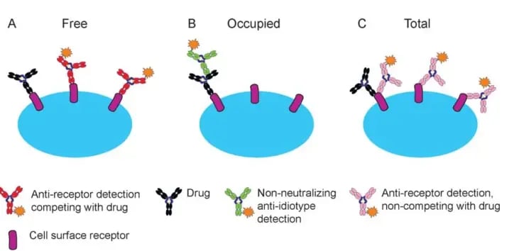
- RO can be measured as free, bound (occupied) or total receptor; the best format depends on the therapeutic and on reagent availability.
Approach
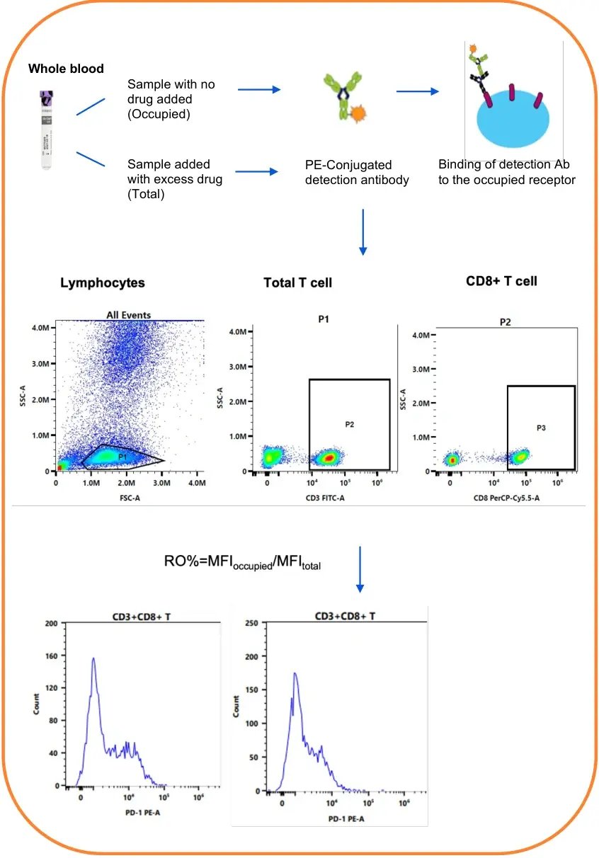
Because the PD-1/anti-PD-1 interaction is dynamic and varies between individuals, and a detection antibody that competes with the drug was not practical, the occupied format was used. Total receptor was measured in parallel by adding excess drug to a second aliquot, which normalizes for baseline expression. Bound drug was detected with a PE-conjugated detection antibody.
Results
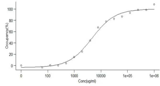
RO was calculated as the ratio of the median fluorescence intensity (MFI) of occupied receptor to that of total receptor. The assay showed good intra- and inter-run precision and a drug-dependent linear range. RO samples were stable for 3 days after blood collection.
| Level | Intra-run CV% | Inter-run CV% |
|---|---|---|
| HQC (~100% RO) | 2.37–7.26 | 7.21 |
| MQC (~50% RO) | 1.19–5.32 | 5.59 |
| LQC (~0% RO) | 0.78–5.31 | 5.87 |
Conclusions
A PD-1 RO assay in the occupied format was established. Measuring total receptor at the same time accounts for baseline expression and reduces bias when receptor levels change during treatment. RO data can also help interpret the effect of ADA on PK and PD in nonclinical and clinical development.
Related services: Receptor occupancy assays · Bioanalysis and biomarkers.
Download the case study
Fill in the short form below and we will email you the PDF (2 pages, 1.3 MB).
To discuss a similar study, email cbs_bd@crystalpharmatech.com or call +1 925-558-5040.